Diabetic Retinopathy

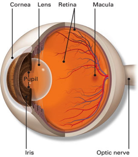
The retina is the inside lining of the back of the eye. It is composed of many layers of light sensitive nerve cells. Light entering the eye is focused on the retina and is converted to electrical impulses which are carried to the brain by the optic nerve. The centre of the retina is called the macula. It is the most sensitive part of the retina and is used for day light and colour vision. This is in contrast to the rest of the retina which is more sensitive in the dark and is mainly used for navigational black and white vision at night.

The retina is supplied by one main artery and one main vein which enter through the optic nerve and branch all over the retina to smaller arteries and veins that cross each other’s like railway tracks.

source – American Academy Ophthalmology

Diabetic retinopathy is a complication of diabetes that causes damage to the blood vessels of the retina—the tissue that lines the back part of the eye, responsible for sharp, detailed vision. Diabetic retinopathy is the most common cause of blindness in the working age group and occurs in more than half of the people who develop diabetes.

Uncontrolled high blood sugar (glucose) level in diabetes causes damage to the blood vessels nourishing the retina. There are different stages of diabetic retinopathy.

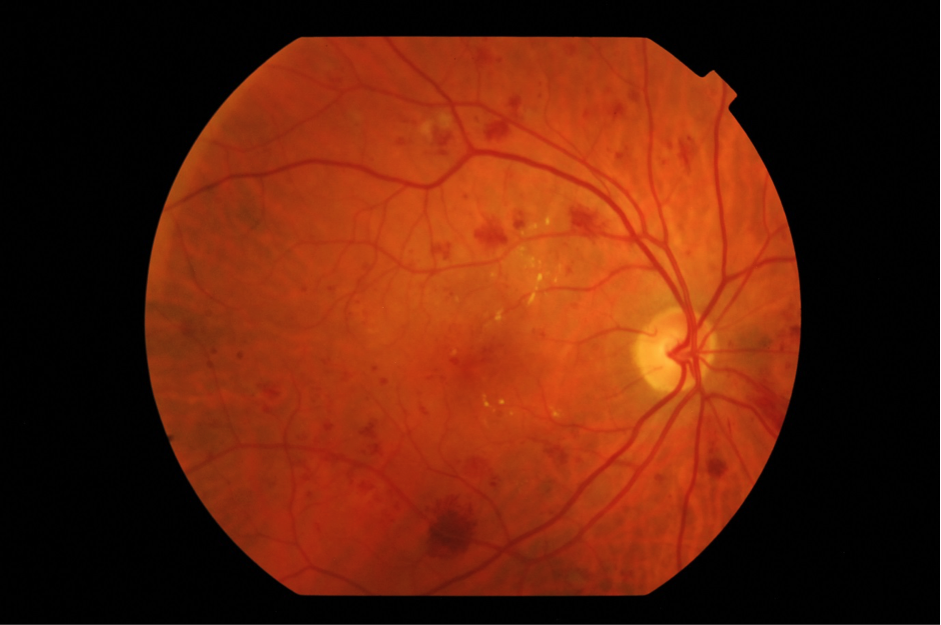
  • The early changes are called mild diabetic retinopathy. Here there retina will have small retinal haemorrhages and dilatation of retina vessels which look like small berries, called microaneurysms. There could also be areas of fluid called oedema or fat deposits called lipid exudates. If the condition progresses with increasing haemorrhages, it is called moderate diabetic retinopathy

Retina photograph showing changes of Moderate Diabetic Retinopathy

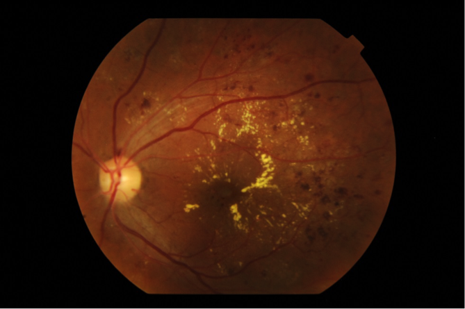
  • Blood vessels can also leak into the centre of the retina (macula) causing swelling. This is called diabetic macular oedema and is the most common cause of reduced vision especially in type 2 diabetes.

Diabetic Macular Oedema

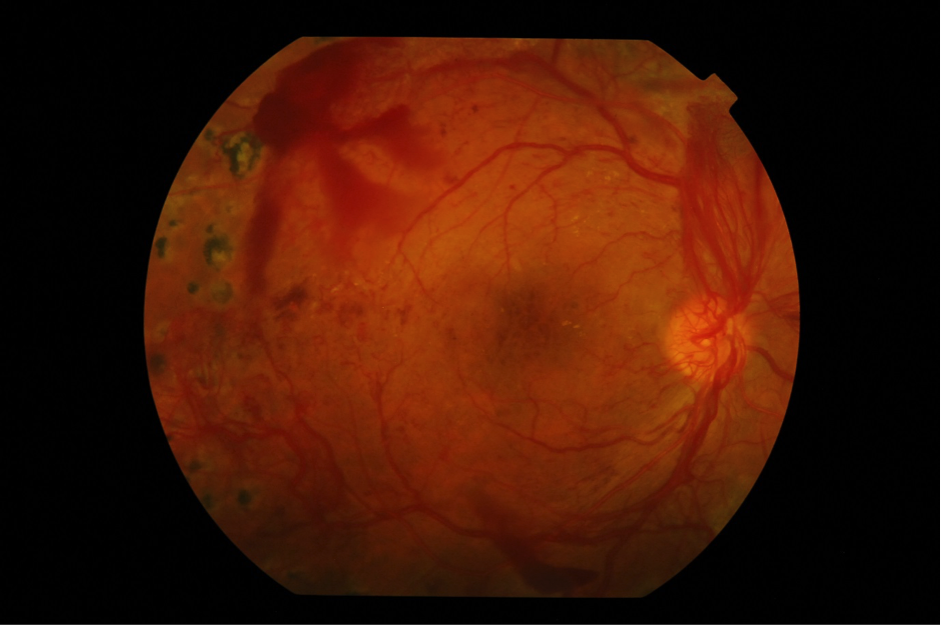
  • If diabetes remained poorly controlled for a long time, circulation to the retina becomes impaired with lack of oxygen and nutrients. This leads to the production of chemicals called “Growth Factors”. These chemicals prompt the development of unhealthy new blood vessels (retina neovascularisation). This stage is called proliferative diabetic retinopathy. The new blood vessels can bleed suddenly into the cavity of the eye (vitreous haemorrhage).

Proliferative Diabetic Retinopathy

  • Eventually scar tissues develop on the surface of the retina that can pull on the retina leading to tractional retinal detachment with severe loss of vision.
  • New vessels can also grow on the iris, in severe cases leading to bleeding in the front part of the eye or causing scarring of the drainage system of the eye. This can lead to high pressure in the eye (rubeotic glaucoma) which can cause pain.

All these changes can eventually lead to severe loss of vision and even blindness if not treated properly.

  • Duration of Diabetes: the longer someone has diabetes, the greater the risk of developing diabetic retinopathy.
  • Poor control of blood sugar levels over time
  • High blood pressure
  • High cholesterol levels
  • Pregnancy

You may not notice any problems with your vision for quite some time even though you may have developed considerable damage to the retina. This highlights the importance of getting your eyes checked regularly if you suffer from diabetes.

Symptoms may include:

  • Blurred vision
  • Distortion; when straight lines appear bent or images are of different sizes.
  • Floaters; that is spots or lines moving in the field of vision
  • Sudden loss of vision
  • Faded colours
  • Eye pain
  • Reading difficulty
  • Dilated retina examination: This is the standard test to check the back of your eye (retina). You will have your pupil dilated with drops so your vision will be blurry for an hour or two after the examination. Our specialist will check your eyes on the microscope (slit lamp) and use a special lens to see the details of the retina. The peripheral retina will be checked with another instrument called an indirect ophthalmoscope. You will also receive a full examination of your eye including the front of the eye (anterior segment), natural lens status to exclude cataract and your eye pressure.
  • Fundus Photography: This is a coloured photo of the retina that helps our specialists to have a record of your eye’s condition at that time and helps them in the long term follow up.
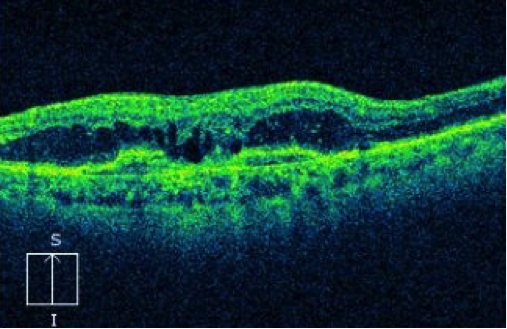
  • OCT (optical coherence tomography); this is a fast scan of retina centre (macula) which shows cross section details of its layer. This test will identify any swelling caused by blood vessel leakage (macular oedema)

OCT of diabetic macular oedema

  • Fundus Fluorescein Angiography (FFA)
  • A fluorescent dye is injected into a vein in the arm or hand. The dye circulates rapidly in the body reaching the retina in a few seconds. A special camera will then take multiple quick pictures which show the dye in the retina circulation clearly. This test helps us to detect swelling and leakage in the macula (macular oedema). It also shows the early budding of new vessels in the peripheral retina (retina neovascularisation) in eyes with proliferative diabetic retinopathy.

FFA of proliferative diabetic retinopathey

  • Ultrasound examination
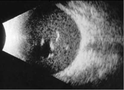
  • If examination of the retina is not possible because of obstructing blood in the eye cavity (vitreous haemorrhage) then an ultrasound test is used to exclude retinal detachment behind the blood.

Ultrasound B-scan of vitreous haemorrhage

The best way of saving sight is to avoid the development of diabetic retinopathy.

  • Maintain good control of your blood sugar level and remain under regular review with your family doctor or endocrinologist (diabetes specialist)
  • Keep your blood pressure under control
  • Keep normal cholesterol and blood lipid levels
  • Stop smoking
  • Have an eye check at the time of diabetes diagnosis. If you do not have diabetic retinopathy at the time of diagnosis, then schedule an eye check every year afterwards.

There have been major advances in the treatment of diabetic retinopathy over the last decade. There are now different methods to treat it and we have become more successful in improving the vision of patient’s who suffer with this serious condition. Each patient will be assessed carefully and advised of the best management approach. We discuss each treatment’s benefit and possible side effects so the patient is fully aware of our goals prior to commencing treatment.

  • Anti-VEGF injections

This is now the main approach to treat swelling of the retina (diabetic macular oedema). It involves injection of drugs into the eye cavity (vitreous). These drugs include Avastin, Lucentis and Eylea. They work by blocking the effect of the chemical produced in the eye with diabetic retinopathy called VEGF (vascular endothelial growth factor).This chemical is the cause of blood vessel leakage in macular oedema and the development of new retinal vessels in proliferative diabetic retinopathy.

Anti –VEGF injections are performed at the Norther Eye Centre without having to be admitted to hospital. They are done under local anaesthesia with no pain felt. We have special expertise in treating diabetic retinopathy and stream line your visit efficiently.

Anti VEGF injections need to be repeated regularly every few weeks to stabilise the macula and prevent recurrent vessel leakage

  • Steroid injections

In some cases steroid drugs may be used to treat swelling of the macula (macular oedema). They can be in the form of a suspension or an implant that releases small doses of steroid over a few months. Repeated injections may also be necessary.

  • Laser treatment

Thermal laser has been used to treat diabetic retinopathy for many decades. It is the main treatment for advanced stage proliferative diabetic retinopathy. Laser treatment is available at our centre and is applied promptly to the patient whilst sitting under anaesthetic eye drops. Some patient may require anaesthetic injection if they feel pain. The treatment is applied over 2 to 3 sessions.

Laser may also be used as an added treatment for swelling of the macula (diabetic macular oedema) usually after a course of anti-VEGF injections.

  • Vitrectomy surgery

Surgery may be necessary for some patients with diabetic retinopathy including those with un-resolving vitreous haemorrhage or with diabetic tractional retinal detachment. The surgery is performed at a day surgery centre in Melbourne which has the latest surgical equipment. Dr Saf Bassili is a highly trained vitreoretinal surgeon with many years of experience.

Want to schedule an appointment?

Call us at (+61) 3 9459 5133 or fill in the appointment form...